Overview
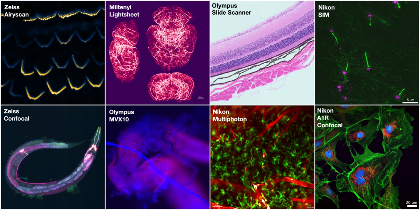
The Microscope Imaging Facility provides access to a wide range of imaging modalities for analysis of biological samples, including multiple systems for brightfield, epifluorescence, confocal, intravital and super resolution imaging. The Nikon and Zeiss confocals are equipped with environmental controls for live cell experiments, and the multiphoton microscope includes anesthesia and physiological monitoring systems for intravital work. Image analysis workstations are available to facility users and these are equipped with software such as Elements, Zen and Imaris to allow quantification of 2D and 3D datasets. Facility staff provide training and support for microscopes and assist users with designing and implementing strategies for analyzing their data.

Equipment
- Miltenyi UltraMicroscope II Light Sheet
- Olympus MVX10 Macroscope
- Nikon A1R Confocal/N-SIM-E*
- Olympus Slide Scanner
- Nikon A1RHD Multiphoton
- Zeiss 710 Confocal w/ Airyscan*
- Nikon Epifluorescence/DIC*
- Zeiss Fluorescent
- Eppendorf Microinjection System
- Zeiss Tissue Culture
Contacts
Director
Neil Billington, PhD | neil.billington@hsc.wvu.edu | (304) 293-0942
Imaging Specialist
Jordan Pascoe | jordan.pascoe@hsc.wvu.edu | (304) 293-2649
Acknowledgements
Please remember to acknowledge the grants that support the Microscope Imaging Facility and any relevant microscopes in your publications, as acknowledgements support our continued operation.
“Imaging experiments were performed in the West Virginia University Microscope Imaging Facility which has been supported by the WVU Cancer Institute and NIH grants P20GM121322 and P20GM144230.”
-
Nikon A1R/SIM: U54GM104942 & P20GM103434
-
Nikon Multiphoton: S10OD026737
-
Zeiss LSM710: P30GM103503 & P20GM103434
-
Zeiss Fluorescent: P20GM103434
-
Olympus VS120 Slide Scanner: P20GM103434
-
Olympus MVX10: P20GM103434
-
Zeiss Tissue Culture: P30GM103488 & P20GM103434
-
Workstations 1 & 2: P20GM103434
